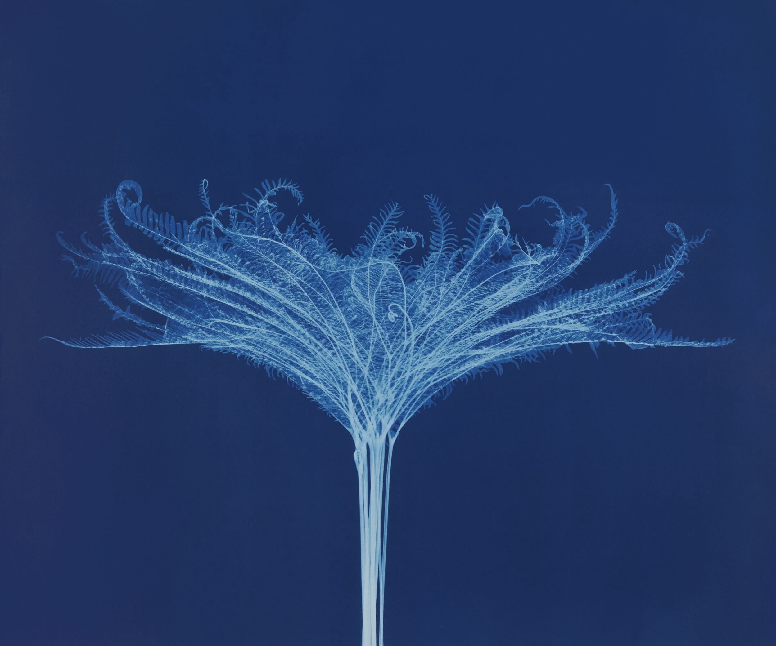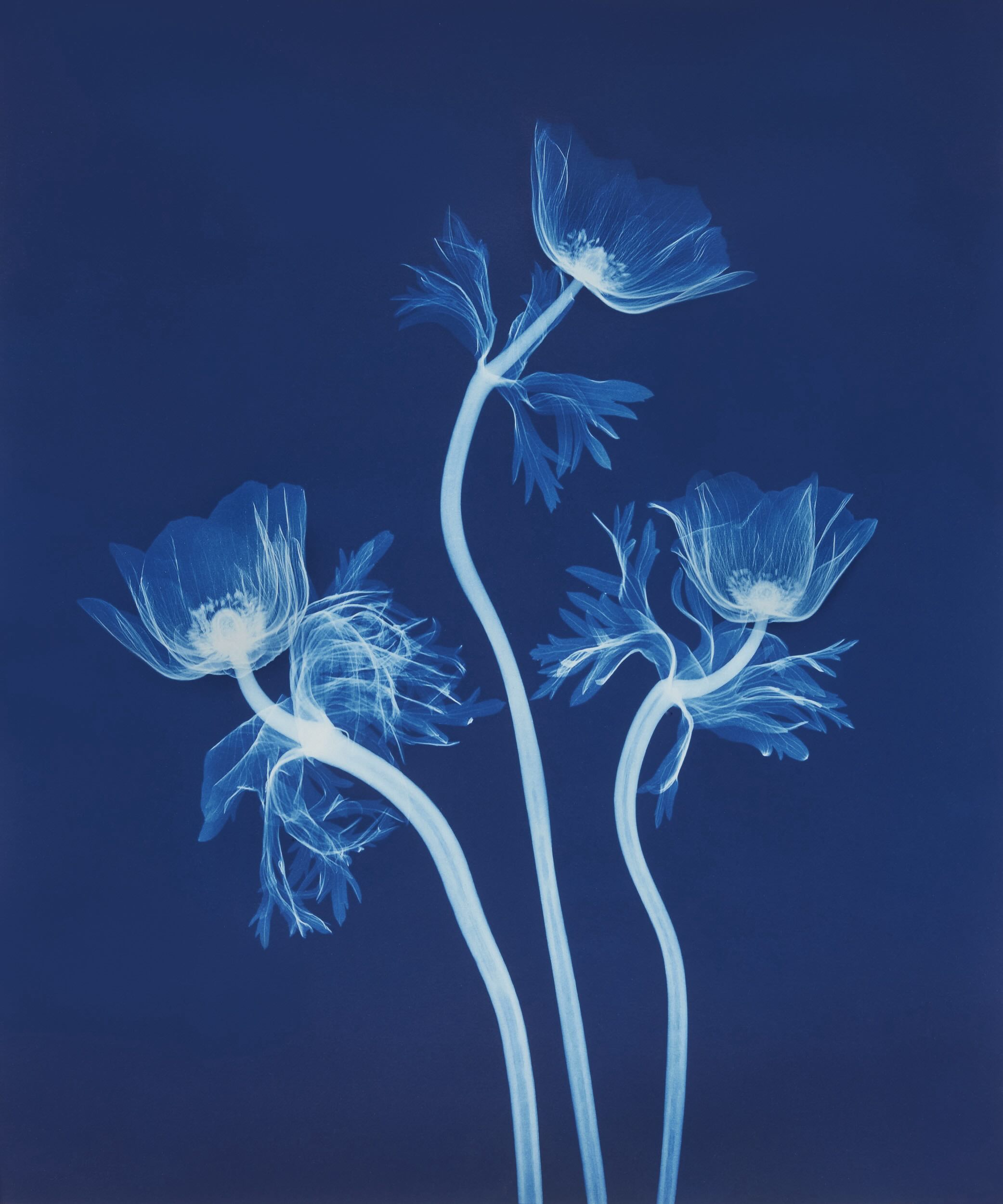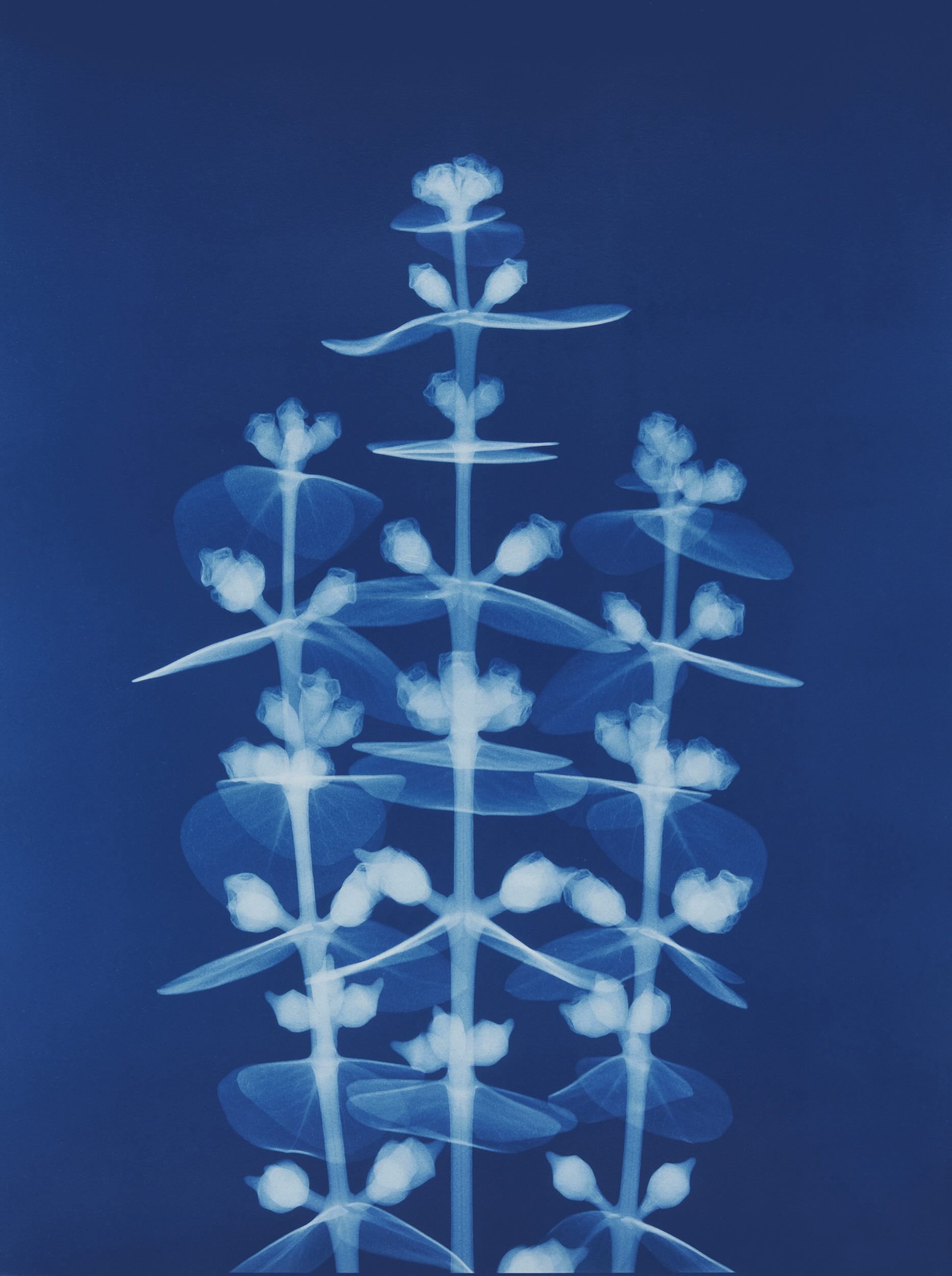
Lilies (detail). 2024. Cyanotype.
My recent show is a culmination of more than 20 years of making X-ray pictures of flowers and plants. Entitled Botanicals in Blue, the exhibition presents a specialized type of camera-less imaging that combines X-rays, discovered in 1896, with one of the first photographic printing processes, cyanotype, invented in the 1840s.
My journey with X-ray imaging started at the Metropolitan Museum of Art where my wife works as a conservator. When I learned that she was using radiography for the examination of art, I was intrigued by the potential to create art with the technique. As a photographer I was always fascinated with experimental imaging, and so after getting permission to perform some tests at the museum I became obsessed with making X-rays of all sorts of objects. Working with other radiologists, I soon used the technique to create commissions for various magazines such Harpers Bazaar, Fortune, and Martha Stewart, and through that work acquired an agent in Paris who landed some high-profile advertising work.
Glass is relatively X-ray opaque, while flowers are very X-ray transparent.
Like so much else in our lives, X-ray imaging is now a digital process, yet I still use traditional photographic development techniques using large sheets of film (17x22”) and develop them in the darkroom. Though it takes much more time to process the images this way, the resolution is higher than its digital equivalent. The size of the film matters because the image formed on the film is in a 1:1 relationship with the subject, much like a shadow of your hand when it is held close to a wall.
Over time I refined my ability how to best translate a plant or flower into an X-ray image. Perspective and angle are important, as is understanding that the front, interior and back of a subject are represented in one plane and seen simultaneously in the image. Also, the X-ray opacity of different materials varies in unexpected ways. For instance, glass is relatively X-ray opaque, because it contains inorganic materials like lead, while flowers are very X-ray transparent, requiring only a minimal amount of power (measured in voltage) to capture.
After the exposure is made and the X-ray film developed, I scan it and work on it digitally. Part of this process includes adjusting the contrast and tonality of the image and then separating the plant from the background. This is a challenging task because the delicate edges often cannot be automatically selected and therefore must be hand-drawn using a digital tablet and pen.

Lotus (in unique frame). Cyanotype. 16 x 20 in / 40.6 x 50.8 cm.
In the past, I printed the X-ray botanicals digitally on fine art paper or transparencies, particularly for large installations. However my newest work is printed with the historic cyanotype process, which uses a light-sensitive iron salt solution brushed onto watercolor paper. Next, an internegative (a second negative of the image made from the original negative) is digitally printed onto acetate at the same size as the final image and placed in contact with the coated paper. When exposed to UV light (either with sunlight or using a UV exposure unit) this coating turns a rich Prussian blue when developed in water, echoing the bluish tint of the original X-ray negative.
For the Santa Fe exhibition I wanted to present the work in a way that would accentuate the physicality of the cyanotype print. Starting with raw lumber, I cut, sanded, and blue-stained pieces of maple to make frames that match the color of the cyanotypes. The result is a handmade object that celebrates the ineffable beauty of plants created by the mysterious qualities of radiation and light. ō
Botanicals in Blue: The X-Ray Cyanotypes of Bryan Whitney
Aurelia Gallery
414 Canyon Rd, Santa Fe, New Mexico
Until June 2

Umbrella Fern. 2024. Cyanotype. 26 x 22 in / 66 x 55.9 cm.

Poppies. 2024. Cyanotype. 26 x 22 in / 66 x 55.9 cm.

Chrysanthemum. Cyanotype. 44 x 44 in / 111.8 x 111.8 cm.

Iris. 2024. Cyanotype. 26 x 22 in / 66 x 55.9 cm.

Eucalyptus. 2024. Cyanotype. 26 x 22 in / 66 x 55.9 cm.

Cactus, 2024. Cyanotype. 26 x 22 in / 66 x 55.9 cm.

Lilies. 2024. Cyanotype. 26 x 22 in / 66 x 55.9 cm.

Rose. 2024. Cyanotype. 26 x 22 in / 66 x 55.9 cm.

BRYAN WHITNEY is a photographer and installation artist. He teaches at the International Center of Photography in New York and exhibits internationally. He lives in New York City.
View Bryan’s X-ray botanical work at www.x-rayphotography.com. His other photographic work can be seen at www.bryanwhitney.com.

Add new comment